Materials Characterisation Laboratory (LCM)
The human and instrumental infrastructure at the LCM is an vital pole for materials research. It is accessible to all researchers in the field. Our mission at the LCM includes the training of students and researchers, the provision of sample analysis services, and the establishment of research collaborations and partnerships.
PRESENTATION
The Laboratory brings together state-of-the-art materials research infrastructures. Major installations of the LCM include several scanning probe imaging techniques (AFM, STM, LFM, MFM, EFM), vibrational microspectroscopy (FT-IR and Raman), ellipsometric spectroscopy and a scanning electron microscope (SEM).
The Department of Chemistry also has modern facilities for the characterization of polymer materials. These equipment are of major interest for materials science and offers a wide range of molecular, nano and micrometric analyzes.
Book a Time Slot Online at the Laboratory
RATES
LCM rates for SPM, micro-Raman & FTIR services
Rates
| Time slot | UdeM & affiliated groups | Other academics | Industrial contracts |
|---|---|---|---|
| Weekdays (Monday to Friday) Autonomous users* | $20 / h | $40 / h | $200 / h |
| Weekdays (Monday to Friday) Service with operator | $55 / h | $90 / h | $290 / h |
| Evening, night and weekends; Autonomous users* | $10 / h | $20 / h | $100 / h |
| Autonomous use* beyond the soft cape treshold (120 h) (anytime) | $5 / h | $10 / h | - |
| Training | $75 | $100 | $150 |
*Allowed user only (after compulsory training)
Advantageous rates (soft cap threshold of 120 h / year)
LCM users can benefit from a preferential rate beyond a flexible threshold of 120 h accumulated during the fiscal year. For example, beyond 120 h, the hourly cost applicable to the use of a technique is 5 $/h for internal academic users (chemistry UdeM, QCAM, GCM, RQMP & CGCC) and $ 10 / hr for other academic users.
Training
To have full access to an LCM system, potential users must attend practical training sessions on the system. The rates are listed below:
- Personal training (one person): $ 75 academic internal users (chemistry UdeM, QCMA, GCM, RQMP & CGCC), $ 100 other academics & $ 150 industry
- Group training (maximum 3 people): $ 50 to $ 100 / additional person
Services and Access Policy
The LCM laboratories are multi-user facilities accessible to the entire scientific community (academic and industrial), given certain user fees. Here is an overview of the services offered by the LCM:
- Training of researchers to the various spectroscopic and microscopic technics available at the LCM, as well as the sustained supervision during the use of the systems.
- Professional sample analysis and characterization services; Assistance in the interpretation of the results obtained.
- Consultation on experimental feasibility : Preliminary exploratory studies to assist some new applicants in assessing the suitability of applying LCM techniques to their specific needs.
- Assistance in developing methods, developing collaborations and research partnerships.
Booking and usage
At least one hour of use is required to validate a reservation.
The maximum number of hours allowed per person, Monday to Friday, is 9 hours per week, spread over one or more days, from 8 am to 5 pm.
However, there are no limitations on the time of use during evenings and weekends (applicable only to certified trained users on the system and for the SPM and FTIR labs).
Reservations are allowed 2 weeks in advance from the current week.
To make a reservation, simply visit the website: LCM Booking Server and follow the instructions.
LABORATORIES
Scanning Probe Microscopy Laboratory
The LCM scanning microscopy unit consists of 7 Atomic Force Microscopes : Bruker (MultiMode 8, Dimension 3100, Dimension ICON FastScan, Dimension ICON in glove box, E-Scope), ANASYS (NanoIR2), WItec (ALPHA 300RS). These multi-function imaging systems combine performance and complementary applications. They are equipped with accessories and additional modules that enhance their operational capacities and broaden their fields of application: different modes and techniques of imaging in air, in liquid and variable temperature.
These equipment of major interest in the fields of nanosciences and nanotechnologies allow the direct nanometric scale characterization of the material surfaces.
Consult the LCM regulations and rates.
Technical Characteristics
- AFM: Atomic force microscopy, in contact Tapping Mode and Peak Force tapping Mode in air and in liquid.
- STM: Scanning Tunneling Microscopy, for measuring the topography of conductive and semiconductive surfaces as well as surface distribution of electron density.
- FMM: Force Modulating Microscopy.
- LFM: Lateral Force Microscopy, probe the mechanical properties (frictional forces) of the surface.
- PIM: Phase Imaging Microscopy, detects chemical heterogeneity at the surface and reveals the different phase domains.
- EFM: Electric Force Microscopy, explores the electrostatic properties at the interface and gives the charge distribution.
- MFM: Magnetic Force Microscopy, image and elucidates the magnetic properties of the surface.
- Scan Asyst: Scan Asyst optimization offers a drastic improvement in lateral resolution in AFM imaging thanks to an improved control of the interaction forces between the tip and the surface combined with an automatic adjustment of the optimization parameters, this improves productivity and reproducibility.
- Peak Force QNM: The PFQNM allows the quantitative mapping of the nano-mechanical properties of surfaces. The nano-mechanical properties such as modulus, adhesion, deformation and dissipation are extracted from each pixel (point-to-sample interaction).
- Peak Force Tuna : PFTUNA for probing conductivity of fragile samples correlated to the quantitative nanomechanical property imaging with PeakForce QNM.
- KPFM : Kelvin Probe Microscopy, allows the mapping of the surface potential with a nm lateral resolution.
- High speed (FastScan): Very high-speed (20 Hz) imaging, allowing to follow dynamic phenomena in real time with a nanometric resolution in a non-damaging way for the sample or the probe.
- Medium: Imaging in air, in liquid medium and at variable temperature.
- Closed-loop scanner: Returns to a defined point (within nm) for force or nanolythographic spectroscopic measurements.
- Open geometry and Sample holder (15x15 sq. in.): Suitable for the coupling of sophisticated experimental setup to AFM and for imaging large parts /components without necessary alterations.
- AFM-Raman: Confocal (2D and 3D) imagery with high spatial resolution (up to 1000 nm) correlated to AFM imaging. Acquisition at a defined point (within a few microns) of Raman spectra.
- SNOM: Scanning Near-field Optical Microscopy, allowing non-destructive optical imaging with a resolution well beyond the diffraction limit, i.e. a 50-100 nm lateral resolution (possibility to work in air or liquid environment) .
- AFM-IR: NanoIR2 allows simultaneous acquisition of topographic images with nanometric spatial resolution coupled to infrared absorption imagery of similar spatial resolution. Acquisition at a defined point (within nm) of infrared spectra over a wide spectral range. Study of the molecular orientation by polarization of the infrared beam.
- NanoTA: Thermal Analysis (phase transitions) localized to the nearest nm.
LCM Vibrational Spectroscopy Laboratory
Raman Spectroscopy
The LCM is equipped with two Raman spectrometers (Renishaw InVia and InVia Reflex) coupled to optical microscopes (Leica) and 4 excitation lasers. This state-of-the-art Raman facility is the Renishaw's most powerful and versatile system. Also, the LCM is equipped with another Raman microscope (Witec Alpha 3000) coupled to two excitation lasers and AFM.
Technical Characteristics
- 5 different wavelengths: 488 nm, 514 nm (>100mW), 532nm, 633 nm, 785 nm
- 3 diffraction gratings: 1800, 1200 et 600 lines/mm
- Conventionnal Raman microspectroscopy and in confocal mode
- Point-by-point Raman spectral mapping
- Raman depth profiling analysis
- Raman global surface imaging at constant wavenumber
- Raman linefocus imaging
- Photoluminescence measurements (PL)
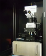
Renishaw InVia Raman Microscope Witec Raman Microscope
Accessories and other options
- Thermostated cell for Raman measurements at variable temperature (-197 to 600 oC)
- Different optical objectives (x5, x20, x50, x40, x100)
- Optical microscope coupled with a video camera for numerical acquisition of structures images
- XYZ motorized stage for sample exploration/fine positioning
- Remote Raman sampling (Raman macro-sampling)
- Possibility for Raman polarization measurements.
Infrared spectroscopy
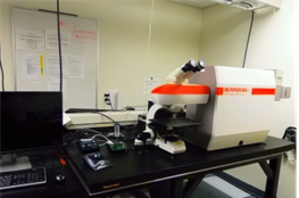
The LCM is equipped with a Stingray FTIR microscope that combines a Digilab FTS7000e Fourier Transform Infrared Spectrometer, an UMA600 IR microscope, and a Lancer detector with a focal plane matrix of 32x32 pixels.
The spectrometer allows the FTIR analysis on a spectral range of 750-4000 cm-1. The Lancer imager is made up of a matrix of 32 by 32 detectors which allows the acquisition of multispectral images at a spatial resolution of 5.5 µm and on an area of 175x175 µm2. The IR system allows FTIR measurements in reflection and transmission modes, and is also equipped with a photoacoustic cell (FTIR-PAS) and a micro-ATR module.
FTIR Microscope Digilab FTS7000e-UMA600
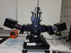
The LCM disposes of a new FTIR-AWI system for performing FTIR measurements with polarized IR light at the air-water interface (AWI) of a Langmuir trough. This system is comprised of a FTIR spectrometer (Bruker Vertex 70) coupled to an external motorized accessory with a Langmuir trough.

FTIR-AWI system, Bruker
LCM Surface Analysis Laboratory
Surface Analysis Laboratory Equipment
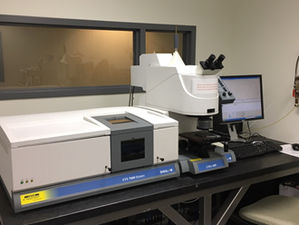
M-2000V Spectroscopic Ellipsometer from J.A. Woollam Co
Ellipsomètre Spectroscopique
INFORMATIONS
Vibrational Spectroscopy
serviceLCM@chimie.umontreal.ca
Fax: 514 343-7586
Scanning Probe Microscopy and Surfaces Analysis
Patricia Moraille (M. Sc.)
Téléphone : 514-34-6111 postes 52008 & 20328
Télécopieur : 514 343-7586
Bureau B-6045 (Campus MIL)
SUPPORT
Support to the LCM:
The LCM receives financial support from the following organizations:
ADDRESS / DELIVERY
| Mailing Address Materials Characterisation Laboratory | Courier Delivery Address Materials Characterisation Laboratory |